Case of the Month
Case of the Month - August 2022

Treatment of Temporomandibular Ankylosis in a Cat with Unilateral Condylectomy

This month's case was a male mixed cat "Lucky", which we think is about 1.5 years old. In the anamnesis taken from the patient's owner, it was learned that the cat was a stray cat taken care of in the school garden. He could not open his mouth fully for about one month and could not eat his food, so he was fed with wet food. The patient’s owner reported that he was brought to our hospital when it was seen that he was weakened because he could not be fed and that his mouth could not be opened when he tried to open it.


    Checking the general condition before starting work is an indispensable examination step affecting each patient's diagnostic and therapeutic approach.

    Habitus: It is related to its environment, is compatible, and has no sign of pain.
    Nutritional status: Poor
    P: 120/beat min, Pulse quality: Normal
    R: 30 /respiration min, Respiration type: Normal, Auscultation: Normal
    Capillary refill time: 1.5 sec, Peripheral pulse: Normal
    Mucous: Slightly pale
    Lymph nodes: Popliteal lymph nodes are normal.
    Abdominal Palpation: Normal

    Special Examination
    His clinical examination observed that the jaw did not open, and a solid mass was palpated at the level of the right ramus mandible.

    Due to the complex anatomy of the skull, four separate radiographic imaging is required for imaging the temporomandibular joint (1,2). It should be examined by taking images in the dorsoventral or ventrodorsal, laterolateral, open mouth, and right or left lateral oblique positions. Radiographic examination was performed under general anaesthesia to properly position the cranial region while radiographs were taken (Figure 1). At the end of the study, left temporomandibular joint ankylosis was diagnosed (Figure 2).

    Figure 1 Radiographic examination of the cranial region. a. Radiograph was taken in the ventrodorsal position. b. Right laterolateral radiography. c. Left laterolateral radiography. D. Radiograph was taken in the right laterolateral oblique position.

    Figure 2 Radiographic examination findings of the cranial region. A. White arrow: Degenerative changes observed in the left temporomandibular joint. B. White arrow: Normal right temporomandibular joint. C. White arrow: Degenerative changes observed in the left temporomandibular joint. D. White arrow: Degenerative changes observed in the left temporomandibular joint.

    The treatment of temporomandibular joint ankylosis is possible with a condylectomy operation. Condylectomy should be performed at the incisura mandibularis, at the base of the condylar process. The osteotomy should be performed in a half-moon shape towards the slightly rostral so that the medial edge of the condylar processes can also be removed (3,4,5,6) (Figure 3).

    Figure 3 Condylectomy line.

    One of the temporomandibular joints was observed normal, and the other was ankylosis. It was predicted that the jaw joint could be mobilised again by performing a condylectomy operation on the joint with ankylosis (Figure 4).

    Figure 4 Intraoperative images of the temporomandibular condylectomy. a. Incision line. b. The temporomandibular joint has undergone degenerative changes. c. Osteotomy is applied to the condyle. D. Removal of the condyle after osteotomy.

    Figure 5: The appearance of the joint after osteotomy and open-mouth X-ray.

    References:

    1. Schwarz T, Weller R, Dickie AM, vd. Imaging of the canine and feline temporomandibular joint: a review. Vet Radiol Ultrasound 2002; 43:85–97.

    2. Ticer J, Spencer CP. Injury of the feline temporomandibular joint: radiographic signs. Vet Radiol 1978; 19:146–56.

    3. Schulz K. Diseases of the joints: Fossum TW, editor. Small animal surgery. 3rd ed. St. Louis, MO: Mosby; 2007. p. 1143–315.

    4. Piermattei DL, Flo GL, DeCamp CE. Fractures and luxations of the mandible and maxilla: Piermattei DL, Flo GL, DeCamp CE, editors. Brinker, Piermattei, and Flo’s handbook of small animal orthopedics and fracture repair. 4th ed. Philadelphia, PA: Elsevier – Saunders; 2006. p. 717–36.

    5. Piermattei DL, Johnson KA. The head: Piermattei DL, Johnson KA, editors. An atlas of surgical approaches to the bones and joints of the dog and cat. 4th ed. Philadelphia, PA: Elsevier – Saunders; 2004. p. 33–45.

    6. Maas CPHJ, Theyse LFH. Temporomandibular joint ankylosis in cats and dogs – a report of 10 cases. Vet Comp Orthop Traumatol 2007;20: 192–7.

    Case of the Month - Janruary 2022

    A CASE OF INTESTINAL LYMPHOMA IN A DOG

    History:
    Our case this month is a 6 years old, male, husky breed dog named "Hatchi". In the history taken from the patient's owner, it was learned that the dog hunched and took a position while defecating for about 3 months, but he had difficulty in defecation and pain (dyschezia), and sometimes he wasn’t able to defecate at all. In addition, when the animal was straining, the presence of an irregular and bleeding mass protruding out of the anus, approximately the size of a walnut, was noticed, and the dog was brought to our hospital.


      Checking the general condition before starting further examinations is an essential step that can affect the diagnostic and therapeutic approach for each patient.

      Habitus: Responsive to surroundings, sometimes shows aggression when in pain.
      Nutritional Status: Bad
      P: 120 beats/min, Pulse quality: Normal
      R: 38 times/min, Respiration type: costo-abdominal, Auscultation: normal
      CRT: 1 sec, Peripheral pulse: normal
      Mucous membranes: Slightly pale
      Lymph nodes: Popliteal lymph nodes are swollen (+2).
      Abdominal palpation: Normal
      Rectal palpation: Awake not possible.

      Special Examination
      On physical examination just cranial to the anus, at the level of 6 o'clock, a painful, bleeding, oval mass with an irregular surface, approximately 3-4 cm in size, narrowing the lumen of the rectum was palpated.

      Our differential diagnosis for the mass detected by clinical examination may be a tumor, granuloma, abscess or hematoma, respectively. As a tumor was suspected as most likely diagnosis, 3-dimensional (right and left lateral, ventro-dorsal) thoracic radiographs and 2-dimensional (right lateral, ventro-dorsal) abdominal radiographs should always be taken for metastasis screening.

      This protocol was applied to Hatchi and no abnormality was observed on direct radiographs.

      An irregularly outlined cyst with hypoechoic content, measuring 1.7 x 1.3 cm, was detected in the right lobe of the prostate.

      Both testicles were of normal size, moderately echoic and homogeneous. A hypoechoic, oval shaped mass of approximately 1 x 1.5 cm with clear margins was also observed in the left testis.

      The remaining abdominal organs were considered normal.

      A: Appearance of the urinary bladder (UB) neck and a hypoechoic cyst (*) in the right lobe of the prostate. There is an irregularly outlined hypoechoic zone in the dorsal part of the prostate (yellow ★) B: Measurement of the cyst in the right lobe of the prostate, C: Hypoechoic mass (yellow ★) in the dorsal aspect of the prostate and its margins to the prostate parenchyma (arrowheads). D: Hypoechoic mass in the left testis and normal right testis.

      On ultrasound the urinary bladder was moderately filled, the content was anechoic, and the wall was thin. The prostate was moderately echoic and homogeneous, but its dimensions could not be clearly determined due to an irregularly shaped hypoechoic structure dorsally. This area was separated from the remaining prostate tissue by an irregular but distinct border. An irregularly outlined cyst with hypoechoic content, measuring 1.7 x 1.3 cm, was detected in the right lobe of the prostate.

      Both testicles were of normal size, moderately echoic and homogeneous. A hypoechoic, oval shaped mass of approximately 1 x 1.5 cm with clear margins was also observed in the left testis.

      The remaining abdominal organs were considered normal.

      A: Appearance of the urinary bladder (UB) neck and a hypoechoic cyst (*) in the right lobe of the prostate. There is an irregularly outlined hypoechoic zone in the dorsal part of the prostate (yellow ★) B: Measurement of the cyst in the right lobe of the prostate, C: Hypoechoic mass (yellow ★) in the dorsal aspect of the prostate and its margins to the prostate parenchyma (arrowheads). D: Hypoechoic mass in the left testis and normal right testis.

      • Prostatic cyst/abscess and hypoechoic area associated with the prostate (leukotic infiltration?).

      • Hypoechoic mass in the left testis (testicular neoplasia: may be sertoli cell tumor, leydig cell tumor or seminoma)

      The mass was digitally palpated from the rectum under general anaesthesia and its relationship with the prostate was investigated. It was determined that the hypoechoic area in the prostate was the mass in the rectum. It was not fully understood whether the rectal mass and the prostate were attached or originated from the prostate (dynamic examination: video). Punch biopsy with a rectal approach was performed for histopathological examination.

      Lymphoma is one of the most common types of tumors in dogs, and the gastrointestinal (GI) tract is the most common extranodal site. In most cases of GI lymphoma, the GI tract is the primary site of disease, but in some cases it is also observed secondary (metastasis). Diagnosis of intestinal lymphoma can be difficult. Histopathology and molecular clonality evaluation are required for definitive results. In accordance with recent studies, the average survival time was determined as 424 days in patients who were treated. It has been observed that survival time increases if the disease responds to treatment (medical/operative).

      According to the results of the pathological diagnosis, Hatchi was given an operation appointment, but the patient did not come.

      References:

      1. Couto KM, Moore PF, Zwingenberger AL, Willcox JL, Skorupski KA. Clinical characteristics and outcome in dogs with small cell T-cell intestinal lymphoma. Vet Comp Oncol. 2018 Sep;16(3):337-343. doi: 10.1111/vco.12384. Epub 2018 Jan 11. PMID: 29322604; PMCID: PMC6041184.

      2. Rassnick KM, Moore AS, Collister KE, Northrup NC, Kristal O, Chretin JD, et al. Efficacy of combination chemotherapy for treatment of gastrointestinal lymphoma in dogs. J Vet Intern Med. 2009 Mar;23(2):317–22.

      3. Sogame N, Risbon R, Burgess KE. Intestinal lymphoma in dogs: 84 cases (1997-2012). J Am Vet Med Assoc. 2018 Feb 15;252(4):440-447. doi: 10.2460/javma.252.4.440. PMID: 29393741.

      In the examined biopsy material atypical neoplastic cells with prominent, round shaped, hyperchromatic nuclei and moderate cytoplasm as well as many apoptotic cells and occasionally atypical mitoses were observed in the intestinal submucosa and between glandular cells. Based on these findings intestinal lymphoma was diagnosed.

      Case of the Month - October 2021

      History

      This month's case is a 4-month-old male Yorkshire terrier dog named “Odin”. Information obtained from the patient owner, revealed that the dog had been vaccinated in a private veterinary clinic. Later complaints of moaning and pain were observed after the vaccination, and symptomatic treatment was initiated in form of painkillers in the same clinic. The dog responded to the treatment for 2 days, but the symptoms recurred. The dog was referred with signs of incoordination to our hospital after the symptoms worsened during the next 2 weeks.


        Checking the general condition before starting work is an essential examination step that can change the diagnostic and therapeutic approach for each patient.

        Habitus: Depression, inability to keep the head still, tremors and weakness in the front extremities were observed.
        Nutritional status: Good
        P: 120 beats/min, Pulse quality: Normal
        R: 38 breaths/min, Respiration type: Costoabdominal, Auscultation: Normal
        Capillary refill time: 1s, Peripheral pulse: Strong
        Mucous membranes: Pale
        Lymph nodes: Normal
        Abdominal Palpation: Normal
        Rectal palpation: Not done

        Neurological Examination:
        In this step, basic functions of the nervous system are checked.
        There were signs of ataxia, incoordination, and pain on palpation of the cranial neck region.
        Cranial nerves: Pupillary light reflex: Delayed, Threat response: Normal, Corneal-Palpebral Reflex: Normal, Nystagmus: None, Labial reflex: Normal.
        Deep pain sensation: Normal, Proprioception: Delayed, Muscle tone: Increased, Wheel barrowing: Negative, Bounce: Negative.

        Odin's blood was taken and a haemogram test was performed.

        Bloodwork did not reveal any evidence that could explain the clinical signs.

        In order to determine the localization of the lesion according to the neurological examination findings, radiographic examination of the head and neck region was performed under general anaesthesia for proper positioning of the cervical vertebrae.

        Did you see the lesion?

        Figure 1: Lateral radiographs taken in neutral position (left) and with mild flexion (right).

        Figure 2: To compare Odin's condition, notice the normal alignment of the vertebrae on the lateral radiograph taken from the neck of a healthy 5-year-old Yorkshire terrier (Left). Odin’s lesion is indicated by the red arrow (right).

        Interpretation of radiographic imaging showed a misalignment between the first and second cervical vertebrae and a significant increase in the distance between both vertebrae. The atlas was displaced dorsally creating also a gap between atlas and occiput.

        👍 Congratulations, correct diagnosis.

        The atlantoaxial joint consists of the first 2 cervical (C) vertebrae, the atlas or C1, and the axis or C2. There are 5 ligaments that provide the stability of the atlantoaxial joint; these are transversal, apical, dorsal atlantoaxial and 2 alar ligaments. Atlantoaxial subluxation (AAS) occurs with abnormal movement of the joint. AAS is most commonly caused by congenital anomalies and is often seen in young, small or miniature breed dogs. Congenital anomalies seen in AAS are aplasia or hypoplasia of the dens, absence or weakness of the supporting ligaments, incomplete ossification of the atlas, block vertebrae, and dorsal angulation of the dens. Abnormal movement of the atlantoaxial joint causes dorsal subluxation of the axis and compression of the spinal cord. Clinical manifestations vary according to the degree of spinal cord compression and range from cervical pain to tetraplegia, respiratory paralysis and death. Traumatic atlantoaxial subluxation (TAAS) occurs when cervical spine trauma leads to instability of the atlantoaxial joint, either from C1-C2 fractures, damage to supporting ligaments, or both (de Lahunta, 2009; Hansen, 2019). Best diagnostic method is computed tomography, which would enable three dimensional evaluation of the case. Treatment methods can be conservative (neck supported bandage + movement restriction) or operative (dorsal/ventral vertebral cerclage, stability with pin, screw, plate and suture materials). The prognosis is always guarded and varies according to the clinical symptoms of the patient and treatment (de Lahunta, 2009; Stalin, 2015).


        The possibility of surviving with neck support was evaluated. However, due to the severity and localization of the lesion, it was decided that this method would not be useful, considering that sufficient stability could not be achieved.

        Due to the severity of the lesion, the prognosis of the operative treatment was guarded and the operative method was not considered appropriate.

        👎 Sorry, misdiagnosed!

        Together with Odin's owner, it was decided to euthanize him before his symptoms further worsened.
         
        Further Reading
        de Lahunta, A and Glass, EN. de Lahunta’s Veterinary Neuroanatomy and Clinical Neurology. Elsevier Health Sciences, 2009.
        Hansen, Sonya C., et al. "Traumatic atlantoaxial subluxation in dogs: 8 cases (2009–2016)." Journal of Veterinary Emergency and Critical Care 29.3 (2019): 301-308.
        Stalin, C., et al. "A review of canine atlantoaxial joint subluxation." Veterinary and Comparative Orthopaedics and Traumatology 28.01 (2015): 1-8.

        Case of the Month - May 2021

        Anamnez

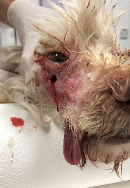
        A 13-year-old male Terrier dog ('Caramel') was presented to our hospital with complaints of difficulty and irritability while eating and bilateral wounds under his eyes for 3 years. It was learned that the wounds did not heal despite treatment and recurred at 4-month intervals.


          Checking the general condition before starting work is an essential examination step that can change the diagnostic and therapeutic approach for each patient.


          Habitus: Alert (related to its environment)
          Nutritional status: good
          P: 100 / min, pulse quality: normal
          R: 30 / min, respiratory costoabdominal, auscultation: vesicular sounds
          Capillary refill time: 2 sec, peripheral pulse:
          Mucosal color: rose-pink
          Lymph nodes: normal
          Abdominal palpation: normal
          Rectal palpation: normal

          Oral examination:
          On intraoral examination under sedation, it was observed that there was no problem related to teeth and gingiva.

          When the wounds were probed, it was determined that the wound had reached the maxillary bone.

          Ophthalmic examination:
          No physical examination findings affecting the eyeball and accessory organs were detected. Examination of the deep layers of the bulbus was not considered necessary.

          Complete blood count (CBC) was performed and leukocytosis (granulocytosis) was detected.

          Did you see the lesion?

          A radiolucent (black) halo indicating bone resorption was observed around the roots of both maxillary 4th premolars on oblique views of the right and left quadrants of the maxilla and mandible. In the light of these findings, it was suggested that the source of the fistula was the inflammation of the tooth roots.


          👍 Well done, correct diagnosis!


          Previous conservative treatment (without a full diagnosis) had been tried unsuccessfully by other vets.
          There is no conservative treatment of carnasial fistula.

          Under general anesthesia, both teeth were extracted operatively.
          After the operation, antibiotics and oral antiseptics were used, and an Elizabethan collar was attached to his head. A few days of soft food feeding was recommended.


          Intraoral connection of the fistula seen intra operatively.

          Post-operative radiographs show that both maxillary P4s were completely extracted without residue.

          👎 Sorry, misdiagnosed!

          On the 15th day after the operation, the sutures of Caramel were removed, the mouth is healed, the fistula wounds are about to close.


          Post-operative 30th day.


          The owner of Caramel stated that he is fine and happier and that he has regained his previous health. As Near East University Faculty of Veterinary Medicine Animal Hospital, we wish him "get well soon, Caramel".

          Authors: Assoc. Prof. Dr. Serkan SAYINER ve Assist. Prof. Dr. Çağrı GÜLTEKİN